Innovative medical ultrasound system design
introduction
Due to its safety, low cost and real-time performance, ultrasound imaging is an important medical imaging method. The traditional ultrasonic imaging system uses the frequency of 2 ~ 15MHz, the accuracy can be accurate to millimeters. They are widely used to monitor the fetus and diagnose diseases of the internal organs of the human body such as the heart, liver, gallbladder, spleen, pancreas, kidney, and bladder.
Due to the large number of channels and large signal processing requirements of ultrasound systems, traditional console-type ultrasound systems have dominated medical ultrasound applications for more than 20 years. The elderly population, increasing healthcare costs and the demands of emerging economies have led to a sharp increase in the demand for innovative medical solutions.
Mature semiconductor technology with higher performance and lower price (for example: digital signal processor (DSP)) not only greatly promotes the development of medical imaging equipment, but also promotes the miniaturization of medical ultrasound imaging systems. In addition, reducing the size of the system does not mean a reduction in performance. Miniaturized ultrasound systems (ie portable ultrasound systems) can achieve the same performance as traditional console-type ultrasound systems. Current portable ultrasound systems can provide better imaging quality to help doctors make accurate and timely diagnosis.
Therefore, portable systems are playing an increasingly important role in applications such as timely trauma diagnosis and emergency and treatment. As more and more ultrasound equipment manufacturers are committed to developing portable ultrasound systems, only those who can launch products more quickly can gain more market share. Ultrasonic analog front end (AFE) and small size, high-performance DSP are all required by ultrasonic equipment manufacturers. More importantly, ultrasonic equipment manufacturers are desperately in need of a design that can be shared with various systems to minimize their development cycle time and accelerate the time to market.
Ultrasonic system structure
Ultrasonic systems vary depending on their function and performance. For example, 3D, 4D and harmonic imaging modes are usually used for high-end systems, while only 2D B-mode imaging and spectral Doppler may be used for some low-end systems. The functional differences mainly depend on the digital backend. High-end ultrasound systems require more and faster computing power, which requires high-end DSPs with near real-time signal processing.
Obviously, it is very difficult to implement shared signal processing units between high-end portable systems. However, without considering different performance requirements, ultrasonic systems usually have similar receive channel architectures.
As shown in Figure 1, the receiving analog front end of the ultrasound system is composed of some general modules, such as: low noise amplifier (LNA), time gain control (TGC) amplifier, voltage control amplifier (VCA), programmable gain amplifier (PGA), Low-pass filter and analog-to-digital converter (ADC).
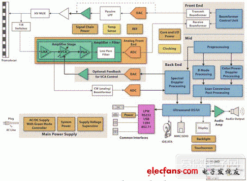
Figure 1 Ultrasonic system structure diagram
In either case, the performance of AFE will greatly affect the performance of the entire system. Therefore, as long as there are AFE products that can meet different performance requirements in a pin-to-pin compatible package, the AFE design can be standardized and reused in various systems. This standardization can be easily achieved in low-end and middle-end systems, which do not require special analog signal conditioning. However, at present, most AFE products cannot meet the needs of ultrasound manufacturers.
Therefore, we must choose some separate chips to meet the different performance requirements of pocket and console systems. For example, although the high power consumption of console-type systems can be tolerated, lower noise must be achieved, and vice versa, and must be redesigned. Now, there are some new AFE devices on the market, for example: TI's AFE5805, which allows ultrasound manufacturers standard AFE design. These devices with the same external pins are mainly used in various ultrasonic systems from portable to console type. Pin-to-pin compatibility means that ultrasound equipment manufacturers can not only design innovative products, but also greatly save costs and accelerate the time to market.
Analog front-end features and system performance
Designing an ultrasound system is a complex task, and every characteristic of AFE can affect the performance of the entire system. The ability to balance various parameters for each system category is undoubtedly an art. For portable ultrasound systems, power consumption is a critical consideration. Low power consumption means that limited battery power can be used for longer working hours. Although these performance degradations are usually within the acceptable range of portable (low-end) systems, they will affect other parameters such as input signal range, input equivalent noise, and harmonic distortion.
In addition to power consumption, AFE noise is the second issue that ultrasonic system designers need to consider. The amplitude of the received signal from the ultrasonic transmitter may range from 10uVPP to 1VPP [1]. The smaller the signal that can be detected, the higher the sensitivity of the system. Both input equivalent current and input equivalent voltage noise will affect the system sensitivity. Generally, we choose the noise parameter of 0.7 nV / rt (Hz) ~ 1.5 nV / rt (Hz) (RTI) for high-end to low-end systems. It has been verified in real systems that these noise parameters are sufficient to produce high-quality images.
Given the input equivalent current noise and the noise from the transmit / receive (T / R) switch, we can even use a lower noise amplifier, but we will not see a significant improvement in the quality of the final ultrasound image. In addition to input equivalent voltage noise, flicker noise (ie 1 / f noise) is also a very important consideration in imaging applications. In continuous wave (CW) mode with mixing, the low-frequency noise spectrum shifts to the carrier frequency, thereby reducing the signal-to-noise ratio (SNR) at the relevant frequency. Due to its wide operating frequency, we will prefer amplifiers with white noise performance.
In some ultrasonic applications, the gain control range plays an important role in obtaining the dynamic range of the image. When VCA has a higher gain control range, the final image has a wider dynamic range, thereby achieving better image quality. Combined with the SNR of the ADC, the dynamic range of the system can be calculated by the following formula:
Dynamic range = SNR + gain control range (Equation 1)
For example, a system that includes a 12-bit, 70dB SNR and 40dB gain control range VCA can achieve a dynamic range of 110dB. In other words, in view of the 0.7dB / cmMHz attenuation coefficient, 10cm imaging depth and 7.5MHz sensor in the human body, the calculation formula for the 105dB dynamic range is 10 * 2 * 0.7 * 7.5. In existing ultrasound systems, 10-15MHz probes are often used to image small parts of the body. Therefore, we usually need a dynamic range above 100dB.
From a system design perspective, an AFE with a larger gain control range is the preferred solution. In addition, an AFE with a higher overall gain is an additional requirement for detecting small signals and compensating for the insertion loss caused by other circuits (for example, the insertion loss of passive high-order filters). Amplifier saturation and overload recovery are also important system parameters. It is more valuable to evaluate and determine these two parameters together than to perform them individually. Basically, the ideal input signal range of an amplifier is limited by its linear output voltage (ie, power supply voltage) and gain:
Therefore, lower gain and higher supply voltage are very beneficial to this parameter. However, lower gain will reduce the input equivalent voltage noise, while higher power supply voltage will increase the total power consumption, so a compromise method must be adopted. We often choose 200 ~ 400mVPP parameters for some portable and mid-range systems. Ultrasonic amplifier saturation is usually caused by large signals reflected by high-voltage pulse leakage or near-surface objects with large changes in acoustic impedance. Specific examples include surface tissues or bones with little clinical information.
In most cases, the loss of information in these areas may not affect the clinical diagnosis. However, if the amplifier cannot be recovered in time, important information will be lost. AFE's fast overload recovery ensures that the ultrasound system can capture as much valuable information as possible. The number of ADC clock cycles can be used to determine the overload recovery time of the AFE, and the load recovery time of one clock cycle is the ideal time.
Another effect of amplifier saturation is the increase in harmonic distortion. Due to the use of common contrast agents, more and more systems (even portable systems) require lower second harmonic distortion throughout the system to ensure successful harmonic imaging. Generally, the harmonic signals received by the transmitter can be as high as 40dB, which is lower than the basic signal, according to the different combinations of contrast agent's acoustic properties, transmitter voltage, and tissue characteristics. Therefore, the HD2 of the amplifier should be less than 40dBc. This allows the system to obtain satisfactory harmonic images.
In addition, due to high HD2, artificial Doppler frequency may appear. In some clinics, this man-made phenomenon will affect the accurate diagnosis. In the final Doppler image, the artificial Doppler moving frequency contributes to the directional separation of the system. Some literature [see bibliography 2 and 3] shows that for CW and PW Doppler systems, directional separation of 45-50dB is sufficient. Considering the above factors, when HD2 is lower than 40dBc, we should specify the linear input range of AFE.
Interference that affects the accuracy of the image is another parameter of the ultrasound system that needs to be considered. Depending on the distance, frequency, design, material, etc. of the transmitter, the ultrasonic system is mainly caused by the array transmitter arranged in the order of –30 ~ –35dBc. Generally speaking, the interference between IC and PCB is much lower than –35dBc. Therefore, the interference of the circuit will not reduce the performance of the system.
Dog Trainer,Patpet Dog Training Collar,Petainer Dog Training Collar,Pet Resolve Dog Training Collar
Elite-tek Electronics Ltd , https://www.aetertek.ca